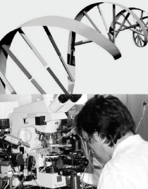
Münster – The enzymes gelatinase A/matrix metalloproteinase-2 (MMP-2) and gelatinase B/MMP-9 are essential for induction of neuroinflammatory symptoms in experimental autoimmune encephalomyelitis (EAE), a mouse model of multiple sclerosis (MS). In the absence of these enzymes, the disease does not develop. SFB128 scientists of Prof. Dr. Lydia Sorokin’s group, therefore, investigated the cellular sources and relative contributions of MMP-2 and MMP-9 to disease at early stages of EAE induction. They demonstrated that MMP-9 from an immune cell source is required in EAE for initial infiltration of leukocytes into the central nervous system and that MMP-9 activity is a reliable marker of leukocyte penetration of the blood-brain barrier.
The neuroscientists then developed a molecular imaging method to visualize MMP activity in the brain using fluorescent- and radioactive-labeled MMP inhibitors (MMPis).
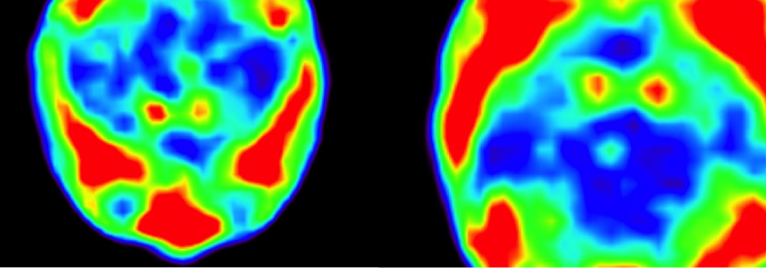
By using radioactive MMP ligand in EAE animals the Muenster neuroscientists produced positron emission tomography (PET) images of MMP activity in patients with MS.
In contrast to traditional T1-gadolinium contrast-enhanced MRI, MMPi-PET enabled tracking of MMP activity as a unique feature of early lesions and ongoing leukocyte infiltration.
MMPi-PET therefore allows monitoring of the early steps of MS development and provides sensitive, noninvasive means of following lesion formation and resolution in murine EAE and human MS, the neuroscientists conclude. Their work was part of the SFB projects B03 and Z02.